Home » Without Label » Anatomy Of Chest - Rotation of 3D skeleton.ribs,chest,anatomy,human,medical ... - Structures of the heart such as the right ventricle (3), intraventricular septum (4), left ventricular free wall (5) and papillary muscles (arrow) are clearly seen.
Anatomy Of Chest - Rotation of 3D skeleton.ribs,chest,anatomy,human,medical ... - Structures of the heart such as the right ventricle (3), intraventricular septum (4), left ventricular free wall (5) and papillary muscles (arrow) are clearly seen.
Anatomy Of Chest - Rotation of 3D skeleton.ribs,chest,anatomy,human,medical ... - Structures of the heart such as the right ventricle (3), intraventricular septum (4), left ventricular free wall (5) and papillary muscles (arrow) are clearly seen.. Organs & structures of the chest heart. Intravenous contrast is seen in the left ventricle (1) and descending aorta (2). Chest a man's chest — like the rest of his body — is covered with skin that has two layers. Related posts of anatomy of the chest area skull of human anatomy body. The chest wall is comprised of skin, fat, muscles, and the thoracic skeleton.
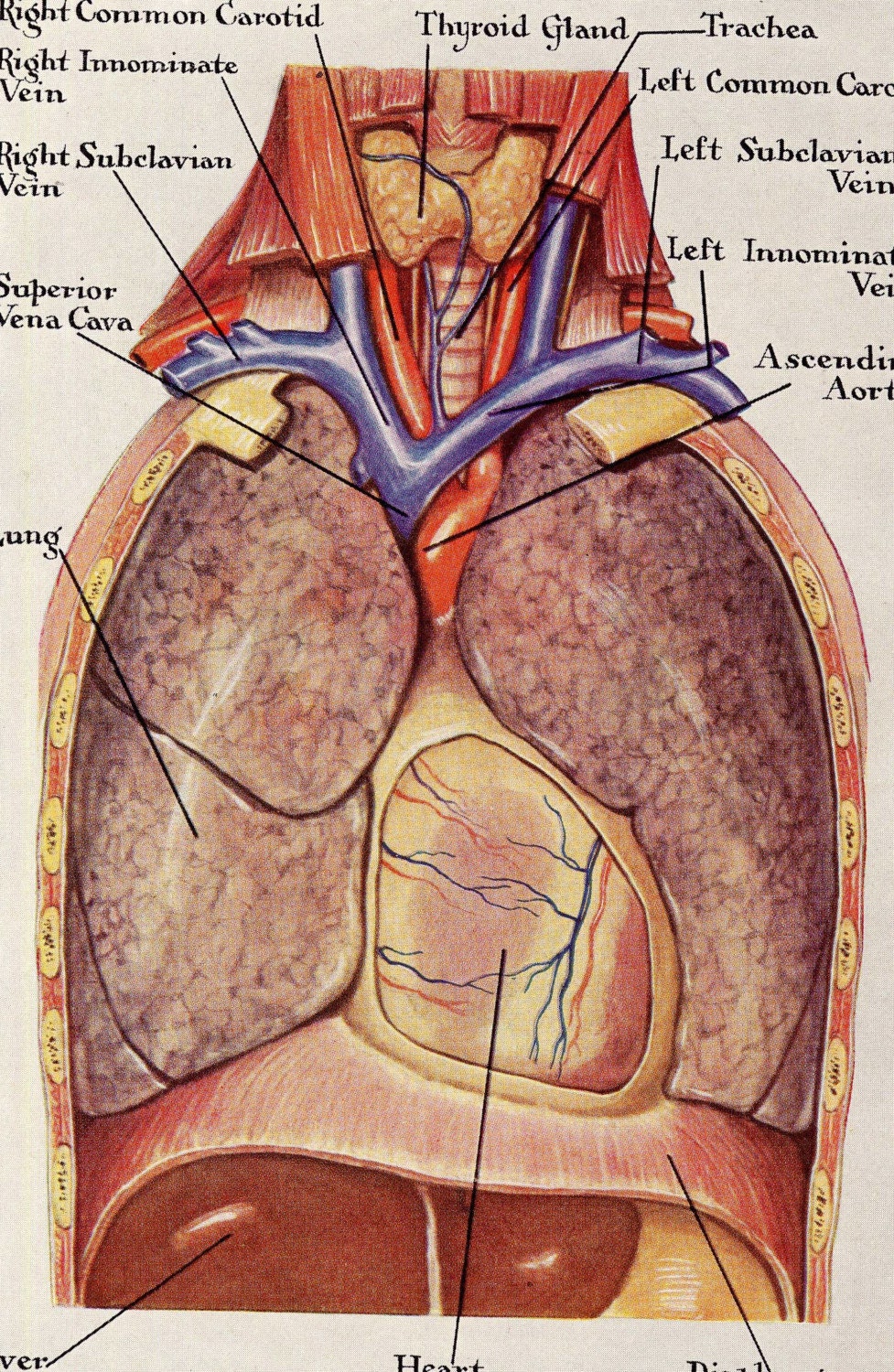
The chest or thorax is the region between the neck and diaphragm that encloses organs, such as the heart, lungs, esophagus, trachea, and thoracic diaphragm. The circulatory system does most of its work. The outer layer of the pericardium surrounds the roots of your heart's major blood vessels and is attached by. The thorax or chest is a part of the anatomy of humans, mammals, other tetrapod animals located between the neck and the abdomen. Anatomy of stomach 12 photos of the anatomy of stomach anatomy of gastric glands, anatomy of stomach and spleen, anatomy of stomach emedicine, anatomy of the stomach area female, parts of stomach ppt, human anatomy, anatomy of gastric glands, anatomy of stomach and spleen, anatomy of stomach emedicine, anatomy of the stomach area.
Human Anatomy Lungs Respiratory System Vintage Medical from img1.etsystatic.com First i'll do an intro to the different organs and structures in the chest, and then i'll go over some images showing their locations. Skandalakis chest wall embryogenesis the muscles of the chest develop from the somites found in the mesoderm. Where is the sternum found. The chest anatomy includes the pectoralis major, pectoralis minor and the serratus anterior. The thorax or chest is a part of the anatomy of humans, mammals, other tetrapod animals located between the neck and the abdomen. Your heart is located between your lungs in the middle of your chest, behind and slightly to the left of your breastbone (sternum). In insects, crustaceans, and the extinct trilobites, the thorax is one of the three main divisions of the creature's body, each of which is in turn composed of multiple segments. Organs & structures of the chest heart.
Structures of the heart such as the right ventricle (3), intraventricular septum (4), left ventricular free wall (5) and papillary muscles (arrow) are clearly seen.
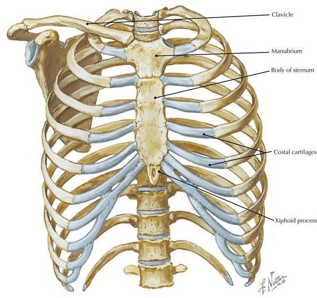
The circulatory system does most of its work. Swensen fund for innovation in teaching. The clinical anatomy of the respiratory tract starts at the external nares or nose. The pectoralis major and the pectoralis minor, known collectively as your pecs. The epidermis is the outermost layer that provides a protective, waterproof seal over the body. Anatomy of stomach 12 photos of the anatomy of stomach anatomy of gastric glands, anatomy of stomach and spleen, anatomy of stomach emedicine, anatomy of the stomach area female, parts of stomach ppt, human anatomy, anatomy of gastric glands, anatomy of stomach and spleen, anatomy of stomach emedicine, anatomy of the stomach area. It is important to remember the position and orientation of the heart when placing a stethoscope on the chest of a patient and listening for heart sounds, and also when looking at images taken from a midsagittal perspective. Anatomy of the chest and the lungs: In insects, crustaceans, and the extinct trilobites, the thorax is one of the three main divisions of the creature's body, each of which is in turn composed of multiple segments. The chest is the area of origin for many of the body's systems as it houses organs such as the heart, esophagus, trachea, lungs, and thoracic diaphragm. About the 6th week, the somites differentiate into the sclerotomes and the dermatomyotomes. It is not unusual to have areas of bruising or erosion to the dorsal tip of the rostal plane. Your sternum is a flat bone in the middle of your chest that protects the organs of your torso from injury.
Front view of muscle anatomy of male chest and torso featuring major muscular groups including the sternocostal, rectus abdominis, pectoralis major, serratus anterior and latissimus dorsi. Related posts of anatomy of the chest anatomy of stomach. Anatomy of the chest and stomach, human anatomy, anatomy of the chest and stomach. The pig has a large horn plate, perforated by the two nares. See chest anatomy stock video clips.
Thorax | Radiology Key from radiologykey.com Thoracic cavity, also called chest cavity, the second largest hollow space of the body. Sternocleidomastoid muscle clavicle and ribs anatomy muscle anatomy chest sternocleidomastoid ribs anatomy chest muscles anatomy thorax rib muscles chest muscles chest anatomy illustration. Intravenous contrast is seen in the left ventricle (1) and descending aorta (2). The epidermis is the outermost layer that provides a protective, waterproof seal over the body. The clinical anatomy of the respiratory tract starts at the external nares or nose. It is not unusual to have areas of bruising or erosion to the dorsal tip of the rostal plane. Anatomy of the chest and stomach, human anatomy, anatomy of the chest and stomach. The chest is made up primarily of two muscles:
Swensen fund for innovation in teaching.
The chest is the area of origin for many of the body's systems as it houses organs such as the heart, esophagus, trachea, lungs, and thoracic diaphragm. This work was supported in part by the kaplow family fund, yale school of medicine. The chest or thorax is the region between the neck and diaphragm that encloses organs, such as the heart, lungs, esophagus, trachea, and thoracic diaphragm. First i'll do an intro to the different organs and structures in the chest, and then i'll go over some images showing their locations. Where is the sternum found. Your sternum is a flat bone in the middle of your chest that protects the organs of your torso from injury. Sternocleidomastoid muscle clavicle and ribs anatomy muscle anatomy chest sternocleidomastoid ribs anatomy chest muscles anatomy thorax rib muscles chest muscles chest anatomy illustration. This page provides an overview of the chest muscle group. Anatomy of the chest and stomach, human anatomy, anatomy of the chest and stomach. The outer layer of the pericardium surrounds the roots of your heart's major blood vessels and is attached by. The tissue adjacent to the aorta is the. Anatomy of the chest and the lungs: The thorax or chest is a part of the anatomy of humans, mammals, other tetrapod animals located between the neck and the abdomen.
It is important to remember the position and orientation of the heart when placing a stethoscope on the chest of a patient and listening for heart sounds, and also when looking at images taken from a midsagittal perspective. The tissue adjacent to the aorta is the. The circulatory system does most of its work. Anatomy of stomach 12 photos of the anatomy of stomach anatomy of gastric glands, anatomy of stomach and spleen, anatomy of stomach emedicine, anatomy of the stomach area female, parts of stomach ppt, human anatomy, anatomy of gastric glands, anatomy of stomach and spleen, anatomy of stomach emedicine, anatomy of the stomach area. Related posts of anatomy of the chest anatomy of stomach.
Surface anatomy of anterior chest wall and lateral chest ... from classconnection.s3.amazonaws.com Anatomy of the thorax, heart, abdomen and pelvis recommended text gray's anatomy for students. The circulatory system does most of its work. Radiology basics of chest ct anatomy with annotated coronal images and scrollable axial images to help medical students and junior doctors learning anatomy. About the 6th week, the somites differentiate into the sclerotomes and the dermatomyotomes. Your heart is located between your lungs in the middle of your chest, behind and slightly to the left of your breastbone (sternum). First i'll do an intro to the different organs and structures in the chest, and then i'll go over some images showing their locations. The chest or thorax is the region between the neck and diaphragm that encloses organs, such as the heart, lungs, esophagus, trachea, and thoracic diaphragm. Skull of human anatomy body 4 photos of the skull of human anatomy body activate javascript android human body applications, how big is a human skull, how many bones are in the skull, how many bones are in your cranium, how many bones are there in skull, neurocranium, skull of human body apps android, what …
This thoracic and pulmonary anatomy tool is especially designed for students of anatomy (medical and paramedical studies).
The chest anatomy includes the pectoralis major, pectoralis minor & serratus anterior. It provides protection to vital organs (eg, heart and major vessels, lungs, liver) and provides stability for movement. This work was supported in part by the kaplow family fund, yale school of medicine. This thoracic and pulmonary anatomy tool is especially designed for students of anatomy (medical and paramedical studies). Learn about each muscle, their locations & functional anatomy. It is enclosed by the ribs, the vertebral column, and the sternum, or breastbone, and is separated from the abdominal cavity (the body's largest hollow space) by a muscular and membranous partition, the diaphragm. This rostal plane or rostrum is used as a rooting and exploration organ and combined with the neck muscles is extremely strong. The pectoralis major and the pectoralis minor, known collectively as your pecs. Computed tomography (ct) of the chest can detect pathology that may not show up on a conventional chest radiograph(1). Skull of human anatomy body 4 photos of the skull of human anatomy body activate javascript android human body applications, how big is a human skull, how many bones are in the skull, how many bones are in your cranium, how many bones are there in skull, neurocranium, skull of human body apps android, what … Swensen fund for innovation in teaching. Applied anatomy of the chest wall and mediastinum petros mirilas michael e. The chest or thorax is the region between the neck and diaphragm that encloses organs, such as the heart, lungs, esophagus, trachea, and thoracic diaphragm.